Abstract
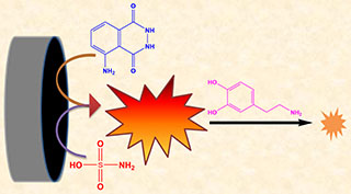
Herein, sulfamic acid (SA) was utilized, for the first time, to enhance significantly the luminol electrochemiluminescence (ECL). With the SA concentration increased from 0.1 μmol·L-1 to 500 μmol·L-1 the ECL intensity increased proportionally. The developed luminol/SA ECL system was employed to detect dopamine (DA) based on its prominent quenching effect. The Stern-Volmer equation of Io/I= 1+Ksv[DA] could be applied to express well the quenching mechanism of DA in the luminol/SA ECL system. The calibration plot showed that the increase in the DA concentration from 0.5 to 20 μmol·L-1 decreased linearly the ECL intensity with a detection limit of 30 nmol·L-1. The luminol/SA ECL system was successfully carried out for DA detection in urine real sample by employing the standard addition method with the excellent recoveries of 103% ~ 105%. Selectivity of the as-developed ECL system was also investigated by using uric acid, ascorbic acid, sugars and amino acids. The results indicated that the ECL intensities changed negligibly in the presence of other substances.
Graphical Abstract
Keywords
electrochemiluminescence, sulfamic acid, luminol, dopamine
Publication Date
2021-04-28
Online Available Date
2021-03-15
Revised Date
2021-03-08
Received Date
2021-02-02
Recommended Citation
Hailemariam Barkae Tesfaye, Ibrahim Halawa Mohamed, Haile Fereja Tadesse, Addisu Kitte Shimeles, Ma Xian-Gui, Chen Ye-Quan, Xu Guo-Bao.
Luminol/Sulfamic Acid Electrochemiluminescence and Its Application for Dopamine Detection[J]. Journal of Electrochemistry,
2021
,
27(2): 168-176.
DOI: Herein, sulfamic acid (SA) was utilized, for the first time, to enhance significantly the luminol electrochemiluminescence (ECL). With the SA concentration increased from 0.1 μmol·L-1 to 500 μmol·L-1 the ECL intensity increased proportionally. The developed luminol/SA ECL system was employed to detect dopamine (DA) based on its prominent quenching effect. The Stern-Volmer equation of Io/I= 1+Ksv[DA] could be applied to express well the quenching mechanism of DA in the luminol/SA ECL system. The calibration plot showed that the increase in the DA concentration from 0.5 to 20 μmol·L-1 decreased linearly the ECL intensity with a detection limit of 30 nmol·L-1. The luminol/SA ECL system was successfully carried out for DA detection in urine real sample by employing the standard addition method with the excellent recoveries of 103% ~ 105%. Selectivity of the as-developed ECL system was also investigated by using uric acid, ascorbic acid, sugars and amino acids. The results indicated that the ECL intensities changed negligibly in the presence of other substances.
Available at: https://jelectrochem.xmu.edu.cn/journal/vol27/iss2/5
References
[1]
Hanif S, Han S, John P, Gao W Y, Kitte S A, Xu G B. Electrochemiluminescence of luminol-tripropylamine system[J]. Electrochim. Acta, 2016,196:245-251.
doi: 10.1016/j.electacta.2016.02.175
URL
[2]
Liu Z Y, Qi W J, Xu G B. Recent advances in electrochemiluminescence[J]. Chem. Soc. Rev., 2015,44(10):3117-3142.
doi: 10.1039/C5CS00086F
URL
[3]
Richter M M. Electrochemiluminescence (ECL)[J]. Chem. Rev., 2004,104(6):3003-3036.
pmid: 15186186
[4]
Sakura S. Electrochemiluminescence of hydrogen peroxide-luminol at a carbon electrode[J]. Anal. Chim. Acta, 1992,262(1):49-57.
doi: 10.1016/0003-2670(92)80007-T
URL
[5]
Hanif S, Han S, John P, Gao W Y, Kitte S A, Xu G B. Electrochemiluminescence of luminol-tripropylamine system[J]. Electrochim. Acta, 2016,196:245-251.
doi: 10.1016/j.electacta.2016.02.175
URL
[6]
Cao Y L, Yuan R, Chai Y Q, Mao L, Niu H, Liu H J, Zhuo Y. Ultrasensitive luminol electrochemiluminescence for protein detection based on in situ generated hydrogen peroxide as coreactant with glucose oxidase anchored AuNPs@MWCNTs labeling[J]. Biosens. Bioelectron., 2012,31(1):305-309.
doi: 10.1016/j.bios.2011.10.036
URL
[7]
S Shkir M, Riscob B, Ganesh V, et al. Crystal growth, structural, crystalline perfection, optical and mechanical properties of Nd3+ doped sulfamic acid (SA) single crystals [J]. Cryst. Growth, 2013,380:228-235.
doi: 10.1016/j.jcrysgro.2013.06.022
URL
[8]
Freeling F, Scheurer M, Sandholzer A, Armbruster D, Nodler K, Schulz M, Ternes T A, Wick A. Under the radar - Exceptionally high environmental concentrations of the high production volume chemical sulfamic acid in the urban water cycle[J]. Water Res., 2020,175:115706.
doi: 10.1016/j.watres.2020.115706
URL
[9]
Upadhyay N, Pujar M G, George R P, Philip J. Development of a sulfamic acid-based chemical formulation for effective cleaning of modified 9Cr-1Mo steel steam generator tubes[J]. Trans. Indian Inst. Met., 2020,73(2):343-352.
doi: 10.1007/s12666-019-01852-4
URL
[10]
Winum J Y, Scozzafava A, Montero J L, Supuran C T. Sulfamates and their therapeutic potential[J]. Med. Res. Rev., 2005,25(2):186-228.
doi: 10.1002/(ISSN)1098-1128
URL
[11] B. Khalili, M. Rimaz, Tondro T. DFT study of N-substituted sulfamic acid derivatives acidity in aqueous media and gas phase[J]. Sci. Iran., 2014,21:2021-2028.
[12]
Lin K N, Xu J, Dong X, Huo Y L, Yuan D X, Lin H, Zhang Y B. An automated spectrophotometric method for the direct determination of nitrite and nitrate in seawater: Nitrite removal with sulfamic acid before nitrate reduction using the vanadium reduction method[J]. Microchem. J., 2020,158:105272.
doi: 10.1016/j.microc.2020.105272
URL
[13]
Kim D S, Kang E S, Baek S, Choo S S, Chung Y H, Lee D, Min J, Kim T H. Electrochemical detection of dopamine using periodic cylindrical gold nanoelectrode arrays[J]. Sci. Rep., 2018,8(1):14049.
doi: 10.1038/s41598-018-32477-0
URL
[14] Egaña L A, Cuevas R A, Baust T B, Parra L A, Leak R K, Hochendoner S, Peña K, Quiroz M, Hong W C, Dorostkar M M, Janz R, Sitte H H, Torres G E. Physical and functional interaction between the dopamine transporter and the synaptic vesicle protein synaptogyrin-3[J]. J. Neurosci. Res., 2009,29(14):4592-4604.
[15] Stanwood G D. Chapter 9 - Dopamine and Stress[M] //Fink G (editor), Stress: Physiology, Biochemistry, and Pathology, Academic Press, 2019: 105-114.
[16]
Khudaish E A, Al-Ajmi K Y, Al-Harthi S H, Al-Hinai A T. A solid state sensor based polytyramine film modified electrode for the determination of dopamine and ascorbic acid in a moderately acidic solution[J]. J. Electroanal. Chem., 2012,676:27-34.
doi: 10.1016/j.jelechem.2012.04.018
URL
[17]
Colín-Orozco E, Ramírez-Silva M T, Corona-Avendaéo S, Romero-Romo M, Palomar-Pardavé M. Electrochemical quantification of dopamine in the presence of ascorbic acid and uric acid using a simple carbon paste electrode modified with SDS micelles at pH 7[J]. Electrochim. Acta, 2012,85:307-313.
doi: 10.1016/j.electacta.2012.08.081
URL
[18]
Tang L J, Li S, Han F, Liu L Q, Xu L G, Ma W, Kuang H, Li A K, Wang L B, Xu C L. SERS-active Au@Ag nanorod dimers for ultrasensitive dopamine detection[J]. Biosens. Bioelectron., 2015,71:7-12.
doi: 10.1016/j.bios.2015.04.013
URL
[19]
Wei X, Zhang Z D, Wang Z H. A simple dopamine detection method based on fluorescence analysis and dopamine polymerization[J]. Microchem. J., 2019,145:55-58.
doi: 10.1016/j.microc.2018.10.004
URL
[20] Ankireddy S R, Kim J. Selective detection of dopamine in the presence of ascorbic acid via fluorescence quenching of InP/ZnS quantum dots[J]. Int. J. Nanomedicine, 2015,10:113-119.
[21]
Huang H, Bai J, Li J, Lei L L, Zhang W J, Yan S J, Li Y X. Fluorescence detection of dopamine based on the polyphenol oxidase-mimicking enzyme[J]. Anal. Bioanal. Chem., 2020,412(22):5291-5297.
doi: 10.1007/s00216-020-02742-1
pmid: 32564120
[22]
Wu B N, Miao C C, Yu L L, Wang Z Y, Huang C S, Jia N Q. Sensitive electrochemiluminescence sensor based on ordered mesoporous carbon composite film for dopamine[J]. Sens. Actuators B Chem., 2014,195:22-27.
doi: 10.1016/j.snb.2014.01.012
URL
[23]
Stewart A J, Hendry J, Dennany L. Whole blood electrochemiluminescent detection of dopamine[J]. Anal. Chem., 2015,87(23):11847-11853.
doi: 10.1021/acs.analchem.5b03345
URL
[24]
Peng H P, Deng H H, Jian M L, Liu A L, Bai F Q, Lin X H, Chen W. Electrochemiluminescence sensor based on methionine-modified gold nanoclusters for highly sensitive determination of dopamine released by cells[J]. Microchim. Acta, 2017,184(3):735-743.
doi: 10.1007/s00604-016-2058-2
URL
[25]
Kim Y R, Bong S, Kang Y J, Yang Y, Mahajan R K, Kim J S, Kim H. Electrochemical detection of dopamine in the presence of ascorbic acid using graphene modified electrodes[J]. Biosens. Bioelectron., 2010,25(10):2366-2369.
doi: 10.1016/j.bios.2010.02.031
URL
[26]
Ma X Y, Chao M Y, Wang Z X. Electrochemical detection of dopamine in the presence of epinephrine, uric acid and ascorbic acid using a graphene-modified electrode[J]. Anal. Methods, 2012,4(6):1687-1692.
doi: 10.1039/c2ay25040c
URL
[27]
Li Z, Zhang H M, Zha Q B, Zhai C Y, Li W B, Zeng L X, Zhu M S. Photo-electrochemical detection of dopamine in human urine and calf serum based on MIL-101 (Cr)/carbon black[J]. Microchim. Acta, 2020,187(9):526.
doi: 10.1007/s00604-020-04524-z
URL
[28]
Venton B J, Cao Q. Fundamentals of fast-scan cyclic vol-tammetry for dopamine detection[J]. Analyst, 2020,145(4):1158-1168.
doi: 10.1039/C9AN01586H
URL
[29]
Zhao H X, Mu H, Bai Y H, Yu H, Hu Y M. A rapid method for the determination of dopamine in porcine muscle by pre-column derivatization and HPLC with fluorescence detection[J]. J. Pharm. Anal., 2011,1(3):208-212.
doi: 10.1016/j.jpha.2011.04.003
URL
[30]
Rao P S, Rujikarn N, Luber J M, Tyras D H. A specific sensitive HPLC method for determination of plasma dopamine[J]. Chromatographia, 1989,28(5):307-310.
doi: 10.1007/BF02260781
URL
[31]
Wen D, Liu W, Herrmann A K, Haubold D, Holzschuh M, Simon F, Eychmüller A. Simple and sensitive colorimetric detection of dopamine based on assembly of cyclodextrin-modified Au nanoparticles[J]. Small, 2016,12(18):2439-2442.
doi: 10.1002/smll.201503874
URL
[32]
Kong B, Zhu A W, Luo Y P, Tian Y, Yu Y Y, Shi G Y. Sensitive and selective colorimetric visualization of cerebral dopamine based on double molecular recognition[J]. Angew. Chem. Int. Ed., 2011,50(8):1837-1840.
doi: 10.1002/anie.v50.8
URL
[33]
Kaya M, Volkan M. New approach for the surface enhanced resonance raman scattering (SERRS) detection of dopamine at picomolar (pM) levels in the presence of ascorbic acid[J]. Anal. Chem., 2012,84(18):7729-7735.
doi: 10.1021/ac3010428
URL
[34]
Figueiredo M L B, Martin C S, Furini L N, Rubira R J G, Batagin-Neto A, Alessio P, Constantino C J L. Surface-enhanced Raman scattering for dopamine in Ag colloid: Adsorption mechanism and detection in the presence of interfering species[J]. Appl. Surf. Sci., 2020,522:146466.
doi: 10.1016/j.apsusc.2020.146466
URL
[35]
Kitte S A, Wang C, Li S P, Zholudov Y, Qi L M, Li J P, Xu G B. Electrogenerated chemiluminescence of tris(2,2'-bipyridine)ruthenium(II) using N-(3-aminopropyl)diethanolamine as coreactant[J]. Anal. Bioanal. Chem., 2016,408(25):7059-7065.
doi: 10.1007/s00216-016-9409-z
URL
[36]
Hui P, Zhang L, Gao W Y, Zuo H J, Qi L M, Kitte S A, Li Y H, Xu G B. Detection of sodium dehydroacetate by Tris(2,2′-bipyridine)ruthenium(II) electrochemiluminescence[J]. ChemElectroChem., 2017,4(7):1702-1707.
doi: 10.1002/celc.v4.7
URL
[37]
Fereja T H, Wang C, Liu F S, Guan Y R, Xu G B. A high-efficiency cathodic sodium nitroprusside/luminol/H2O2 electrochemiluminescence system in neutral media for the detection of sodium nitroprusside, glucose, and glucose oxidase[J]. Analyst, 2020,145(20):6649-6655.
doi: 10.1039/D0AN01178A
URL
[38]
Bancirova M. Sodium azide as a specific quencher of singlet oxygen during chemiluminescent detection by luminol and Cypridina luciferin analogues[J]. Luminescence, 2011,26(6):685-688.
doi: 10.1002/bio.1296
pmid: 21491580
[39]
Gao W Y, Wang C, Muzyka K, Kitte S A, Li J P, Zhang W, Xu G B. Artemisinin-luminol chemiluminescence for forensic bloodstain detection using a smart phone as a detector[J]. Anal. Chem., 2017,89(11):6160-6165.
doi: 10.1021/acs.analchem.7b01000
URL
[40]
Fereja T H, Kitte S A, Gao W Y, Yuan F, Snizhko D, Qi L M, Nsabimana A, Liu Z Y, Xu G B. Artesunate-luminol chemiluminescence system for the detection of hemin[J]. Talanta, 2019,204:379-385.
doi: 10.1016/j.talanta.2019.06.007
URL
[41]
Buettner G R, Ng C F, Wang M, Rodgers V G J, Schafer F Q. A new paradigm: Manganese superoxide dismutase influences the production of H2O2 in cells and thereby their biological state[J]. Free Radical Biol. Med., 2006,41(8):1338-1350.
doi: 10.1016/j.freeradbiomed.2006.07.015
URL
[42]
Khaket T P, Ahmad R. Biochemical studies on hemoglobin modified with reactive oxygen species (ROS)[J]. Appl. Biochem. Biotechnol., 2011,164(8):1422-1430.
doi: 10.1007/s12010-011-9222-2
URL
[43]
Rowley D, Halliwell B. Formation of hydroxyl radicals from hydrogen peroxide and iron salts by superoxide- and ascorbate-dependent mechanisms: relevance to the pathology of rheumatoid disease[J]. Clin. Sci., 1983,64(6):649-653.
pmid: 6301745
[44] Whitman C L. Titrimetric determination of sulfamic acid[J]. Anal. Methods, 1957,29(11):1684-1685.
[45]
Wahba M E K, El-Enany N, Belal F. Application of the Stern-Volmer equation for studying the spectrofluorimetric quenching reaction of eosin with clindamycin hydrochloride in its pure form and pharmaceutical preparations[J]. Anal. Methods, 2015,7(4):10445-10451.
doi: 10.1039/C3AY42093K
URL
[46]
Gong A Q, Zhu X S, Hu Y Y, Yu S H. A fluorescence spectroscopic study of the interaction between epristeride and bovin serum albumine and its analytical application[J]. Talanta, 2007,73(4):668-673.
doi: 10.1016/j.talanta.2007.04.041
URL
[47]
Parajuli S, Jing X H, Miao W J. Electrogenerated chemiluminescence (ECL) quenching of the Ru(bpy)32+/TPrA system by the explosive TNT[J]. Electrochim. Acta, 2015,180:196-201.
doi: 10.1016/j.electacta.2015.08.107
URL